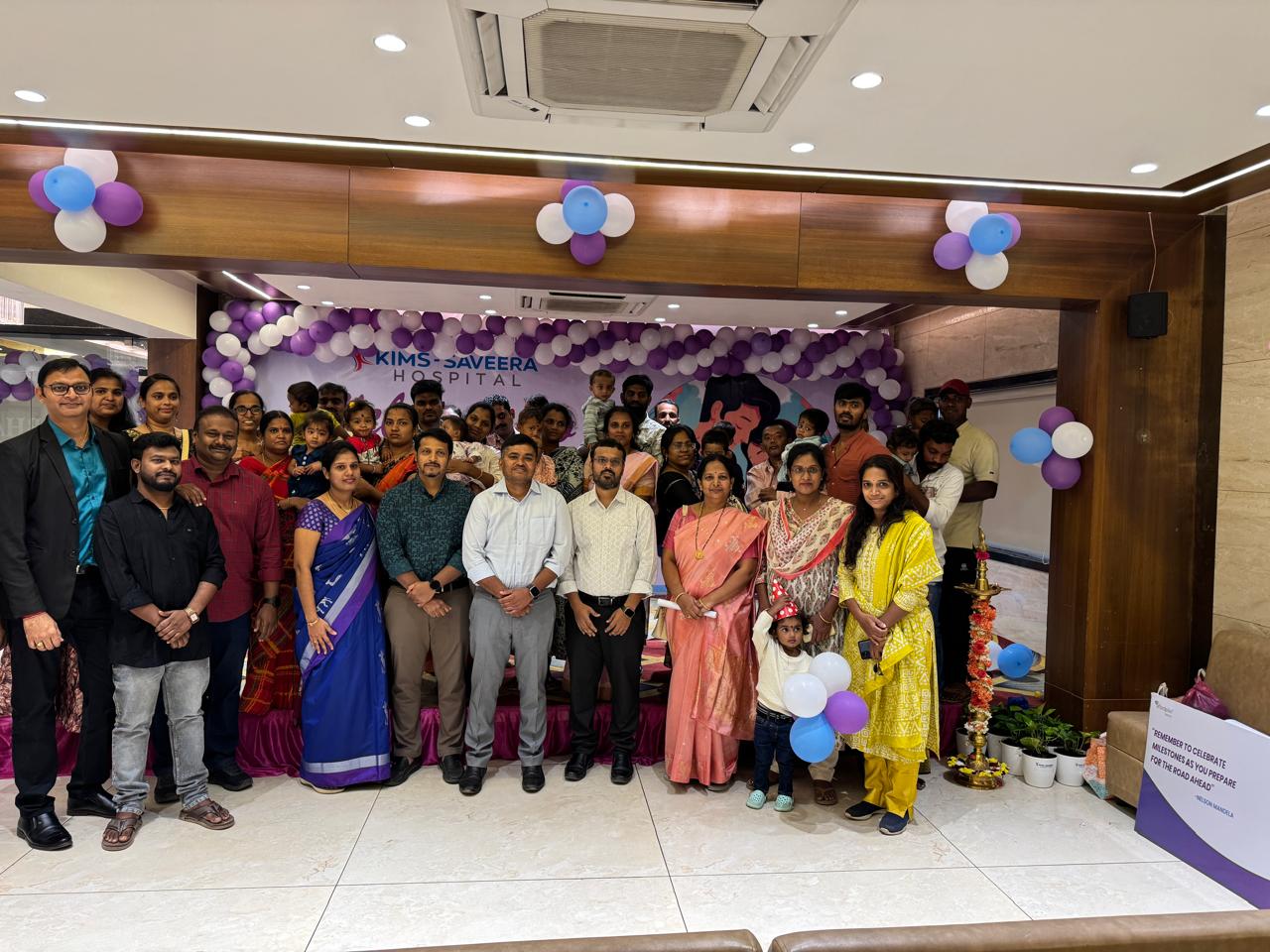
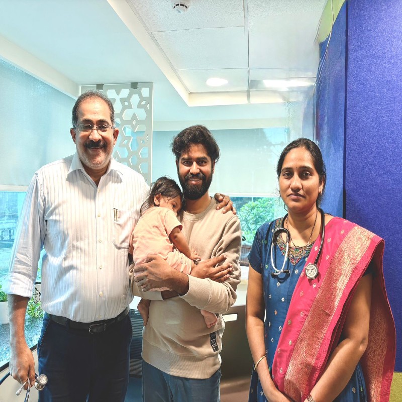
Congenital Heart Defect Awareness week
– February 7th to 14th 2021
Dr. Shanti Priya
Consultant Pediatric Cardiologist
KIMS ICON HOSPITAL
VISAKHAPATNAM
A congenital heart defect (CHD) is a defect in the structure of the heart which is present at birth. Congenital heart defects are the most common type of major birth defects.These anomalies arise when there is a failure of the heart or its major blood vessels to form properly during fetal growth and development.
Considering a birth prevalence of congenital heart disease as 9/1000, the estimated number of children born with congenital heart disease in India is more than 200,000 per year. Of these, about one-fifth are likely to have serious defect, requiring an intervention in the first year of life
They may range from simple holes in the cardiac septum, or narrow valves, to more complex defects like tetralogy of Fallot (ToF), which is a combination of four distinct defects. Most infant deaths from CHD occur within the neonatal period. An infant’s chances of survival are dependent on the severity of the CHD, the time before it is diagnosed, and the success of treatment.
Simple CHD
Among the simple CHD are septal defects. These arise due to holes in the walls of the heart, which cause oxygen-rich and oxygen-poor blood to mix. The children with these septal defects can present with rapid breathing, difficulty feeding and poor weight gain.
Atrial septal defects (ASD) are found between the two upper chambers of the heart, namely, the right and left atria. Many children with ASD often do not have many symptomsand about half of these children will have the ASD close over time on its own.
Ventricular septal defects (VSD) occur in the two lower chambers of the heart (the ventricles) and also allow the mixing of oxygen-deficient and oxygen-rich blood. There is normally a difference in pressures between the left and right ventricles of the heart, with the left being under higher pressure than the right.When there is a hole in the ventricles, the higher pressure from the left ventricle is transmitted to the right ventricle resulting in higher pressure in the lungs and causes damage to the pulmonary vasculature.Smaller VSD, like ASD, may close on their own, but larger ones require intervention. Untreated symptomatic large VSDs can eventually lead to heart failure and death.
Another example of a simple CHD is patent ductus arteriosus (PDA). This condition is rather common and occurs when there is an abnormally persisting communication between the pulmonary artery and the aorta. These two vessels physiologically communicate in utero via the ductus arteriosus, which is vital in the fetal circulation to allow the nonfunctioning lungs to develop.The communication closes usually within three days after birth, but may remain patent in some babies.
Complex CHD
Of the complex CHD, Tetralogy of Fallot (ToF) is the most common. It consists of a combination of four major defects, namely, a VSD, pulmonary stenosis, overriding aorta and hypertrophy of the right ventricle. ToF disrupts the normal oxygenation of blood in the lungs, and it causes oxygen-deficient blood to be pumped to the rest of the body.These children present with cyanosis i.e bluish discoloration of lips, tongue and nails.
Other types of complex CHD:
- Transposition of the great arteries, which is the reversal of the normal anatomical position of the aorta and pulmonary arteryleading to an interruption in blood flow to the lungs or body.
- Total anomalous pulmonary venous connection (TAPVC) causes oxygen-rich blood to enter the wrong chamber, because the veins from the lungs are incorrectly connected
- Epstein’s anomaly, a failure in the tricuspid valve that is usually accompanied by an ASD
- Coarctation of the aorta which is narrowing of this great vessel, and there is a disruption in the free flow of blood to the body.
- Complete atrioventricular canal defect (whole spanning all of the heart’s four chambers)
- Truncus arteriosus occurs when there is one common great arterial trunk as opposed to two separate arteries supplying blood to the body and lungs.
- Surgery is required to correctcomplex CHD.
Treatment
Treatment options vary, depending on the nature and severity of the defect.
Procedures that involve catheterization may be used in combination with surgery. However, such a combination is not always necessary, and these techniques may be done independently according to the nature of the CHD.In addition to these invasive procedures, non-invasive pharmacotherapy may also be used in some instances.
Catheterization
This involves the use of a thin and flexible tube that is inserted into a blood vessel such as the femoral vein, and then threaded backward into the heart. It is a less invasive procedure in comparison to surgery, and as a result it is much better tolerated and enables a quicker recovery.
Catheterization is generally the treatment of choice for simple CHD, such as stenosis of the pulmonary valves or atrial septal defects (ASDs). A small device which is used to close the defect is passed in along the catheter. It may have two small disks to fit, one on each side of the hole. Tissue grows over these disks over a period of months.
When there is stenosis of the pulmonary valve, a catheter is used to insert a balloon that will eventually be inflated in order to open up the valve leaflets. It is subsequently deflated and removed after the valve opening is sufficiently enlarged.
Surgery
If defects cannot be repaired with the help of catheterization, then surgery is necessary. Open-heart surgical procedures involve opening up the chest to gain direct access to the heart. Various procedures are adopted, including placing patches to close defects, repairing or replacing valves, or transplanting various parts of the heart and its great vessels.
Screening:
Fetal echocardiography, a special type of ultrasound scan to examine fetus for abnormalities can provide a relatively fair assessment of majority of fetal heart defects.
Although most new-borns with critical CHD exhibit the symptoms and are identified soon after birth, some are not diagnosed until after they are discharged from the hospital. This makes screening for CHD immediately after birth very important. CHD can be detected through clinical examination and by performing the pulse oximetry test which measures the baby’s pulse and oxygen levels in the blood. A low oxygen level is one of the indicators of CHD.
Early detection of congenital heart disease is of paramount importance to improve the quality of life and prevent morbidity and mortality. Early diagnosis and timely intervention is crucial in appropriate management of children with congenital heart defects.
Covid 19 pandemic and CHD
These groups may be at increased risk for COVID-19: single ventricle patients after Fontan operation, those with chronic cyanosis and depressed ventricular function, individuals with severe pulmonary hypertension, immune-compromised patients (including those who have undergone heart transplantation), infants with unrepaired significant congenital heart disease, and adults with CHD complicated by coronary artery disease or systemic hypertension.
It is recommended that all cardiac medications, including aspirin, anticoagulants, ACE inhibitors, angiotensin receptor blockers, beta blockers, diuretics and antiarrhythmic medications be continued during COVID-19 illness, unless a clear contraindication develops.


